


概要
文書
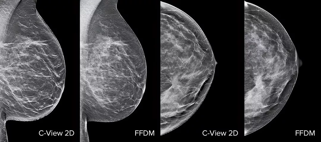
低線量、正確性の高い検査
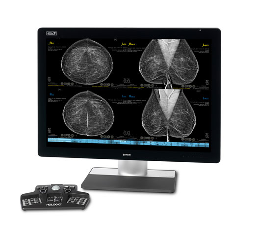
C-View ソフトウェアおよび即座に生成された合成 2D 画像を使用して、乳がん検診のパフォーマンスが向上します1-4。細部が強調され、分析が速くなるだけでなく、患者への放射線量レベルも低減します。C-View 2D 画像は臨床実績があり3,5、トモシンセシス検診検査における FFDM 画像を用いた診断の代替となります。
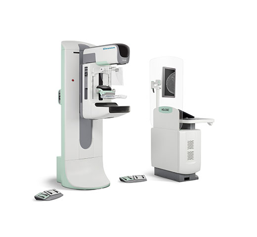
C-View 2D イメージングは、Selenia® Dimensions® システムおよび 3Dimensions® システムの両方で使用できる選択肢です。標準解像度の 3D イメージングにのみ対応しています。高解像度の 3D イメージングには対応していません。
進化した技術で、診断を次のステージへ
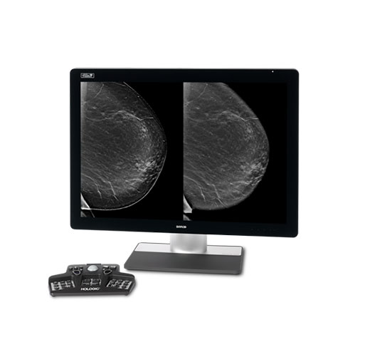
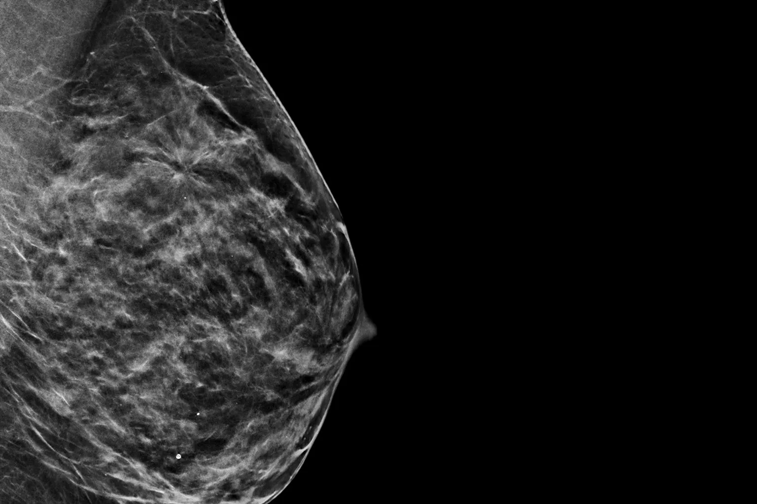
C-View 2D 画像では、微小石灰化でよく見られる構造の歪み、腫瘤性病変、および輝点が、従来の FFDM 2D 画像や断層スライスよりもよく見えます4,6-9。
優れたパフォーマンス
全ての乳房タイプについて、2D マンモグラフィ単独よりも優れた臨床パフォーマンスが得られます4。
リスクの軽減
3.7 秒の超高速スキャン*10,11で再撮影のリスクを軽減します5,7。
より高い正確性
より低線量でより高い正確性**7。
時間の有効活用
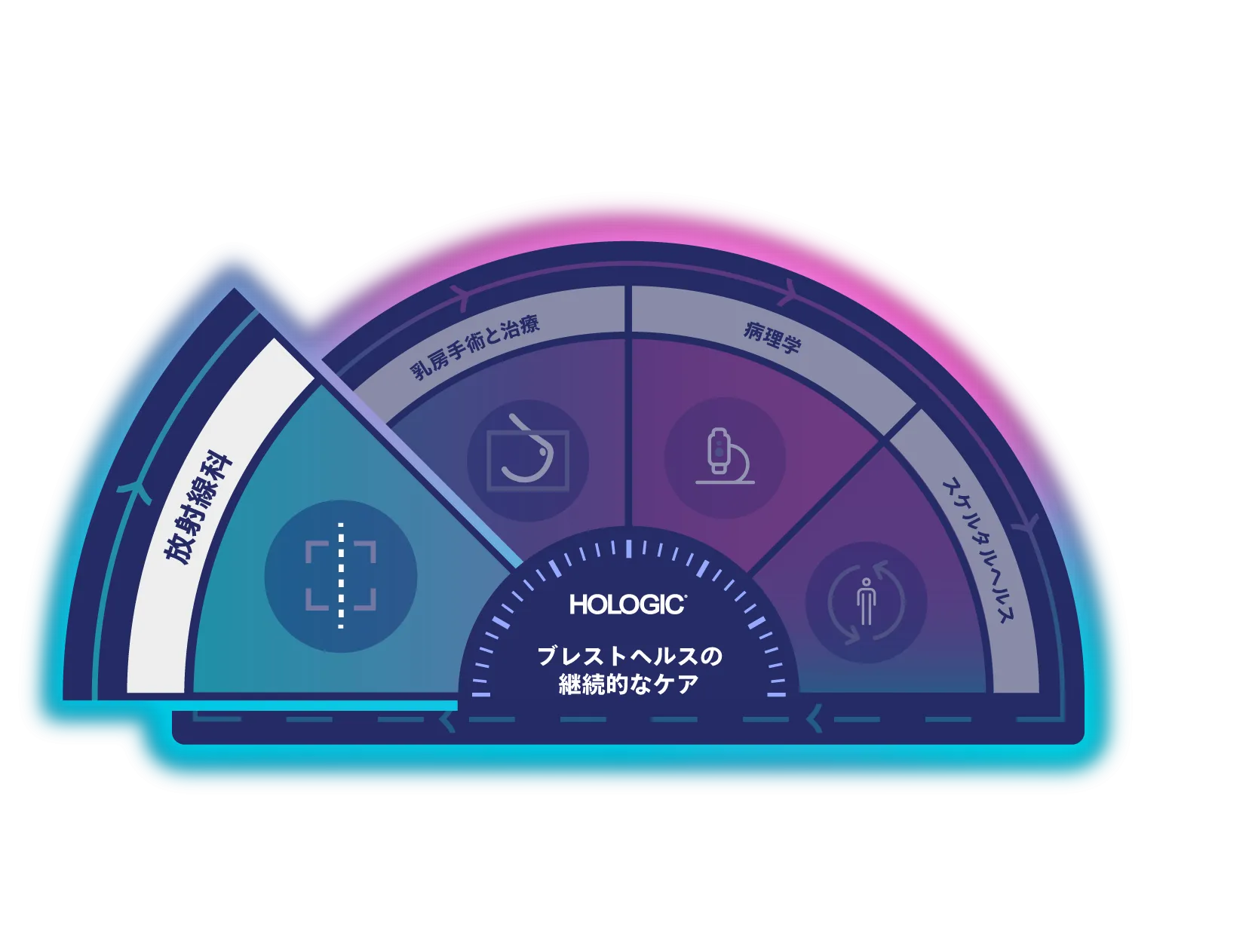
「ブレストヘルスの継続的なケア」は、臨床的信頼性、ワークフローの効率、思いやりのある患者ケアのための統合ソリューションを提供します。より多くの女性の健康増進を支援することが私たちの目的です。
C-View 合成 2D イメージングは、ホロジックの検診および診断ソリューションの一部です。
細部にこそ証拠がある
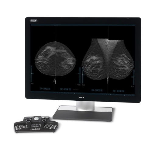
C-View 2D 画像は臨床実績があり3,5、トモシンセシス検診検査における FFDM 画像を用いた診断の代替としてご使用いただけます。これらの画像は、トモシンセシススライスレビューのナビゲーションの補助にもなります。発表された研究によると、低線量 3D マンモグラフィ検査では、2D 単独の場合と比較して、浸潤がんをより早期に発見し、偽陽性の再検査率も低減することが示されています4,5,7。
C-View 2D 画像では、微小石灰化や構築の乱れ、腫瘤性病変、および輝点が、従来の FFDM 2D 画像や断層スライスよりもよく見えます4,6-9。
販売名:デジタル式乳房 X 線撮影装置 Selenia Dimensions
医療機器認証番号:222ABBZX00177000
*他の標準モデルとの比較
** DBT + 合成 2D と DBT + 標準 2D の比較
-
Friedewald SM, Rafferty EA, Rose SL, et al.Breast cancer screening using tomosynthesis in combination with digital mammography.JAMA.2014 Jun 25;311(24):2499-507.
-
Rafferty EA, Durand MA, Conant EF, et al.Breast Cancer Screening Using Tomosynthesis and Digital Mammography in Dense and Nondense Breasts.JAMA.2016;315(16):1784-6.
-
Zeng B, Yu K, Gao L, Zeng X et al.Breast cancer screening using synthesized two-dimensional mammography:A systematic review and meta-analysis, Elsevier Breast 2021 Oct; 59:270–278.
-
Bernardi D, Macaskill P, Pellegrini M, et. al.Breast cancer screening with tomosynthesis (3D mammography) with acquired or synthetic 2D mammography compared with 2D mammography alone (STORM-2): a population-based prospective study.Lancet Oncol.2016 Aug;17(8):1105-13.
-
Skaane P, Bandos A, Eben E, et al.“Two-View Digital Breast Tomosynthesis Screening with Synthetically Reconstructed Projection Images:Comparison with Digital Breast Tomosynthesis with Full-Field Digital Mammographic Images” Radiology.2014 Jun; 271(3):655-63.
-
Durand M, Raghu M, Geisel J, et al.“Synthesized 2D Mammography + Tomosynthesis:Can We See Clearly?”(2015 年 12 月、イリノイ州シカゴで開催された北米放射線学会年次総会で発表された論文)。
-
Zuckerman SP, Conant EF, Keller BM, et al.Implementation of Synthesized Two-dimensional Mammography in a Population-based Digital Breast Tomosynthesis Screening Program.Radiology.2016 Dec;281(3):730-736.
-
Zuley M, Guo B, Catullo V, et al.“Comparison of Two-dimensional Synthesized Mammograms versus Original Digital Mammograms Alone and in Combination with Tomosynthesis Images.”Radiology.2014 Jun;271(3):664-71.Epub 2014 Jan 21.
-
Woo O, Choi G, Shin H, et al.“Comparative Diagnostic Value of Two-dimensional Synthesized Mammogram and Conventional Full-field Digital Mammogram for Evaluation of Breast Cancer”(2015 年 12 月、イリノイ州シカゴで開催された北米放射線学会年次総会で発表されたポスター)。
-
Rocha Garcia AM., Mera Fernandez D. Breast tomosynthesis:State of the art.Radiologia.2019;61(4):274-285
-
Lai Y-C, Ray KM, Mainprize JG, et al.Digital Breast Tomosynthesis:Technique and Common Artifacts.Journal of Breast Imaging 2020;2:615-28.